Oral Biopsy & Lesion
Evaluation in Wylie, TX —
Answers. Not Uncertainty.
A sore that keeps lingering, a white or red patch, a lump you can't explain — the reassuring truth is that most of these turn out to be harmless. But the stakes are too high to guess: caught early, oral cancer carries an 88%+ five-year survival rate, against roughly 38% once it has spread. So Dr. C examines the area thoroughly and biopsies it when there's any question — trading uncertainty for a real answer.
"If there's one thing I want worried patients to hear, it's this: don't sit on it and hope it disappears. The two-week rule exists for a reason. If a sore hasn't healed by then, come let me look. Nine times out of ten it's nothing serious — but on the tenth, finding it early is what makes all the difference."
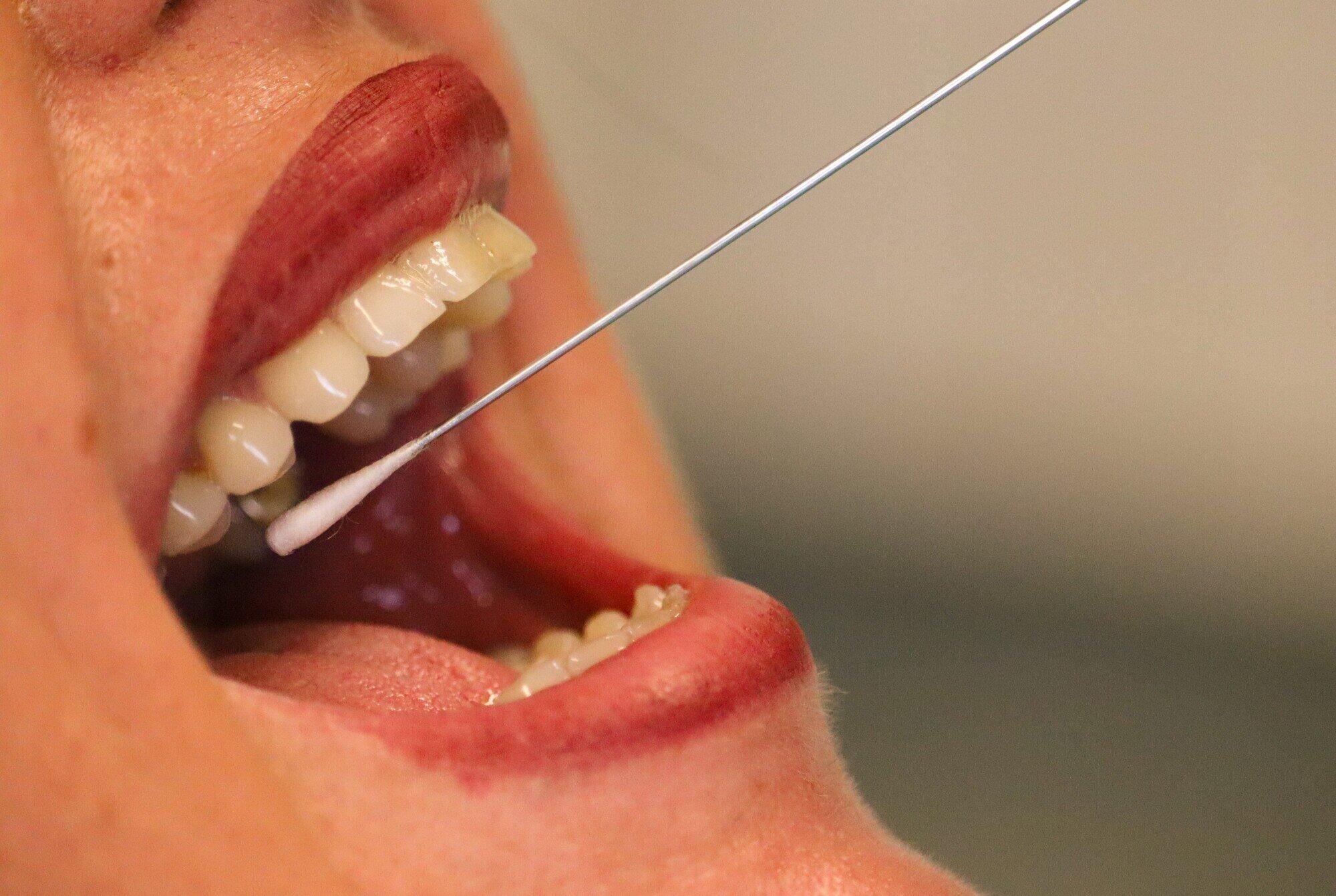
🔬 Book Same-Week EvaluationWarning Signs That Deserve a Prompt Look — Don't Sit On Them
Most changes in the mouth turn out to be harmless. But missing an early oral cancer carries such serious consequences that having any suspicious change checked is simply the smart move. Spot any of the signs below? Give Dr. C a call this week.
Sore That Won't Heal
An ulcer or sore that's still there after 2 weeks is among the clearest red flags. Everyday canker sores and bite injuries close up in 7–14 days; anything that outlasts that window should be looked at by a professional rather than left to "see if it goes away."
White Patch (Leukoplakia)
When a white patch on the gums, tongue, cheeks, or palate won't rub away, it's called leukoplakia. Plenty of cases are benign, yet it's officially classed as a potentially malignant disorder — and only a biopsy can tell whether dysplasia (abnormal cell changes) has set in.
Red Patch (Erythroplakia)
A vivid red, velvety patch on the lining of the mouth with no obvious explanation. Erythroplakia is far more likely to be malignant than a white patch — in the published literature, most biopsied cases already show severe dysplasia or invasive carcinoma. This one shouldn't wait.
Unexplained Lump or Thickening
A fresh lump, a thickened or rough patch anywhere in the mouth, on the lips, or in the throat that sticks around past 2 weeks. If an area simply feels different under your tongue — firmer, rougher — it's worth examining whether or not it actually hurts.
Numbness or Tingling
Lasting numbness, tingling, or a dulled sense of feeling in the mouth, lips, tongue, or face — especially when there's no clear trigger such as recent dental work. When a nerve is affected, it can point to deeper tissue changes that need a closer look.
Pain When Swallowing or Chewing
Ongoing pain, soreness, or a "something's stuck" feeling while chewing, swallowing, or talking that a known dental problem doesn't account for. Trouble swallowing that lingers is reason enough for a prompt evaluation.
Mixed Red-White Patch
Sometimes called erythroleukoplakia or "speckled leukoplakia" — a patch blending red and white areas. That mix is regarded as more dangerous than an all-white patch, which is why a biopsy is the standard response every time.
Voice or Speech Changes
Hoarseness that won't clear or a voice that simply sounds different for no clear reason — especially alongside other mouth symptoms or a tobacco or alcohol history — can signal changes in throat tissue that merit evaluation.
Bite or Denture Fit Changes
When your bite suddenly feels off, or a denture that always fit well turns uncomfortable, and ordinary dental wear doesn't explain it — the cause can be underlying tissue change in the jaw or mucosa that's worth investigating.
⚠️ Why two weeks is the line: Ordinary sores — canker sores (aphthous ulcers), a cheek you accidentally bit — clear on their own inside 7–14 days. Once a lesion passes the two-week mark, don't bank on it healing later. Call Dr. C at (972) 483-4848 and we'll see you that same week. The SEER/NCI numbers leave little doubt: catching it early is what shifts the outcome.
What Is an Oral Biopsy — and Why Is It the Gold Standard?
Only a biopsy can deliver a definitive, under-the-microscope diagnosis of oral tissue. The peer-reviewed literature — including a narrative review from the American Head and Neck Society Cancer Prevention Service — is consistent on the point: biopsy is the universally accepted gold standard for diagnosing suspicious oral lesions.
Oral Biopsy Process — From Lesion to Diagnosis
Why Won't a Visual Exam Alone Settle It?
No clinician, however seasoned, can reliably tell benign tissue from malignant by sight alone. Two lesions that look identical in the chair can be completely different under magnification — one harmless reactive tissue, the other severe dysplasia or early invasive cancer. The microscope simply sees what the naked eye can't.
That's exactly why biopsy stays the gold standard: it yields a real histopathological diagnosis — the actual cellular makeup of the tissue — instead of an educated guess. When something looks suspicious, an impression isn't enough. You're owed a definite answer.
📖 Published evidence (AHNS Cancer Prevention Service, 2025): In a PubMed/MEDLINE narrative review (searched August 31, 2025), the American Head and Neck Society Cancer Prevention Service states plainly that "Biopsy is widely accepted as the gold standard diagnostic method for lesions raising suspicion of malignancy." It notes incisional punch biopsy as reproducible and frequently sufficient on its own, with brush cytology serving as a low-impact, anesthesia-free way to start.
📊 Why early detection is the whole game: SEER (NCI) projects 60,480 new oral cavity and pharynx cancers in 2026. Found while still localized, five-year relative survival reaches 88.7%; after it spreads regionally, that figure falls steeply. Yet only 25.9% are caught at that localized stage — the clearest argument there is for routine screening and not sitting on a suspicious lesion.
📚 Evidence & Sources
NCI SEER Program (2026). SEER Cancer Stat Facts: Oral Cavity and Pharynx Cancer — National Cancer Institute, Bethesda, MD. Reports localized five-year relative survival of 88.7% and an estimated 60,480 new cases in 2026. seer.cancer.gov/statfacts/html/oralcav.html
American Head and Neck Society Cancer Prevention Service (2025). A PubMed/MEDLINE narrative review (searched Aug 31, 2025) backing the claims here: biopsy as the gold standard for suspicious oral lesions, incisional punch biopsy as reproducible and usually sufficient, and brush cytology (OralCDx) as an anesthesia-free first screen.
Published peer-reviewed literature on oral potentially malignant disorders — leukoplakia, erythroplakia, and biopsy technique — shapes the evaluation and management guidance offered here, alongside Dr. C's UCSF training and 20-plus years of oral pathology practice.
Three Types of Oral Biopsy Available at Merry Dental Hub
Which technique Dr. C chooses depends on the lesion's size and location, how suspicious it looks, and what keeps you most comfortable. Every method is done right here in the office — with local anesthesia where it's needed — and the sample goes to an accredited oral pathology lab.
A small spinning brush is pressed against the lesion and rotated to lift a full-thickness sample of cells, which is then smeared onto a glass slide and mailed to the lab. There's no anesthesia, no cut, and no stitches — it doesn't hurt at all. For anyone wary of needles or generally anxious about dental visits, it's an easy first screening step.
Here a representative piece of the suspicious area is taken — and with a larger lesion, the portion that looks most concerning is the part sampled. A punch biopsy uses a small circular cutter (usually 3–6mm); a scalpel biopsy uses a surgical blade. Local anesthesia keeps you comfortable throughout, and the site is closed with sutures. AHNS Cancer Prevention Service guidance calls incisional punch biopsy reproducible and often enough to provide the diagnostic information needed.
This time the whole lesion comes out, so it doubles as both the diagnosis and the treatment in a single sitting — ideal for small, well-defined spots that are most likely benign. Removing all of it gives the pathologist the most tissue to work with and cuts down on sampling error. Local anesthesia and sutures, same as the incisional approach.
What to Expect — From Evaluation to Results
Nothing happens without Dr. C explaining it first. From the evaluation through the biopsy itself, he keeps the communication open, calm, and straightforward at every stage.
Dr. C looks and feels his way through the whole mouth — every mucosal surface, the top, sides, and underside of the tongue, the floor of the mouth, palate, gums, lips, and the back of the throat — and gently checks the lymph nodes in your neck. The lesion is photographed as a baseline, and he reviews your medical history and how long the symptoms have been there.
He lays out what he's seeing in plain terms and recommends the biopsy method that fits the lesion. If starting with brush cytology makes sense, he'll say so; if the picture calls for going straight to an incisional or excisional biopsy, he'll explain the reasoning. You'll know exactly what's about to happen, and what the possible outcomes are, before anyone proceeds.
For an incisional or excisional biopsy, local anesthetic goes precisely around the lesion and the area is fully numb in 2–3 minutes. Dr. C won't start until you tell him you're numb. That little injection is usually the only thing you'll feel the whole time — collecting the tissue itself is painless. Brush cytology skips anesthesia entirely.
The sample is taken with whichever method was chosen — brush, scalpel, or punch — and for incisional or excisional biopsies a few sutures neatly close the site. The specimen drops straight into formalin fixative, gets labeled with your details, and ships out to an accredited oral pathology lab that same day.
Dr. C goes over the results with you face to face — not a context-free phone call, not just a portal notification. He explains the findings in everyday language, hands you a written summary, and spells out what comes next: a monitoring plan if it's benign, or a coordinated referral to an oral medicine specialist or head-and-neck oncologist if dysplasia or cancer shows up. You're never left without a clear path. Should follow-up care touch the nearby teeth, our root canal and cosmetic restoration pages cover the next steps.
After the Biopsy — Recovery Information
Immediately after the procedure
A little soreness as the numbness fades is completely normal, and over-the-counter ibuprofen usually handles it. Once feeling fully returns you can eat and drink as usual — just keep very hot or hard foods off the biopsy side for 2–3 days. Beyond that, no special diet.
Days 2–7 — healing progress
As it heals, the site often turns white or pale yellow — that's normal granulation tissue, not a problem. Expect light soreness for 3–5 days. Starting 24 hours out, gentle salt-water rinses keep things clean. Skip vigorous swishing, resist poking it with your tongue, and try not to chew on that side.
Suture removal (if placed)
If the stitches don't dissolve, a quick 7–10 day follow-up takes care of removing them. Often we use dissolving sutures instead, which need no removal at all. Dr. C tells you which kind you have at the procedure visit.
When to call Dr. C after biopsy
Reach us at (972) 483-4848 if bleeding won't settle after 20 minutes of firm, gentle pressure; if pain ramps up rather than easing after Day 3; if you see signs of infection like growing swelling, spreading redness, or fever; or if the site just looks well off from the normal healing Dr. C described.
"Before I ever share a biopsy result, I tell the patient what our next move will be — because what unsettles people most isn't the result itself, it's not knowing what happens after. Benign or not, there's always a defined path. The one thing I refuse to do is hand someone a worrying result and leave them sitting with a question mark."
What Happens After Biopsy — Your Complete Recovery Timeline
Healing from an oral biopsy is usually quicker and simpler than people brace for. Here's the day-by-day picture of what's normal — and the point at which you should pick up the phone.
Day-by-Day Recovery
First 24 Hours — Most Critical
A bit of oozing or spotting at the site is normal — if it's actively bleeding, bite firmly on gauze for 30–45 minutes. Don't rinse, spit, or use straws; the suction pulls the forming clot loose. Use an external ice pack 20 minutes on, 20 off, to keep swelling down, and stick to soft, cool foods like yogurt, smoothies, and pudding (no straws). Rest with your head propped up. The first 12 hours tend to be the most noticeable, and it improves quickly from there.
Days 2–3 — Soreness Peaks
Soreness usually tops out around Days 2–3 and eases off afterward. Now that the first 24 hours have passed, start gentle saltwater rinses (½ tsp salt in 8oz of warm water) to keep the area clean and help it heal. Over-the-counter ibuprofen (adults, as directed) is normally plenty for the discomfort. A whitish or yellowish look at the site is healthy fibrin tissue — not an infection.
Days 4–7 — Improving, Sutures Present
By Day 4–5 the pain has usually dropped off sharply. Dissolving sutures (from an incisional or excisional biopsy) start loosening and may partly break down over Days 7–14. Keep up the saltwater rinses 2–3 times a day, and steer clear of hard, crunchy, spicy, or piping-hot foods for the whole first week. Most people are back to their normal routine by Day 3–4.
Days 7–14 — Results & Follow-Up
The lab generally turns the tissue around in 7–14 business days. Dr. C reaches out to you directly to walk through the results and next steps — by phone first if that comes before your follow-up. Any sutures are dissolved or removed by this point, and he checks the site to confirm it's healing as it should.
What's Normal — What's Not
✓ Normal & Expected
🚨 Call (972) 483-4848 If You Notice:
🍽️ What to Eat — First Week
✓ Eat freely
✗ Avoid
🦷 Does an oral biopsy hurt? Most patients call it mild — closer to a routine filling than an extraction. The numbing injection is the high point of discomfort; the biopsy itself is pain-free, with maybe 2–3 days of light soreness to follow. Nervous about it? Ask about our sedation options →
Oral Cancer Risk Factors — Who Should Be Screened Regularly
Oral cancer doesn't discriminate — it can show up in anyone. That said, some factors clearly raise the odds. If one or more of the items below describes you, making oral cancer screening at Merry Dental Hub a routine is especially worthwhile.
Tobacco Use
Every form counts — cigarettes, cigars, pipes, plus chewing tobacco, snuff, and other smokeless products, which raise risk too. The longer and heavier the habit, the more it stacks up. Tobacco remains the single biggest preventable cause of oral cancer. Regular oral exams matter most for tobacco users →
Heavy Alcohol Use
On its own, heavy or regular drinking pushes risk upward. Pair it with tobacco and it's especially hazardous — the two together multiply the danger well beyond what either does alone.
HPV Infection
Human papillomavirus — HPV-16 in particular — is behind a growing share of oropharyngeal cancers, often in younger adults who never smoked or drank. HPV-linked oral cancers are now the fastest-rising category of cases across Western countries.
Sun Exposure (Lip)
Years of UV exposure specifically raises the risk of lip cancer, the most common site for sun-driven oral cancers. Outdoor workers, athletes, and anyone with heavy lifetime sun exposure should keep a close eye on changes to the lips.
Age Over 40
Risk climbs with age, and historically most diagnoses have landed in people past 40. But with HPV-related oropharyngeal cancers climbing among younger adults, screening now matters across a far broader age range than it used to.
Male Sex
Oral cancer strikes men about twice as often as women. That gap is shrinking as tobacco and alcohol habits shift across groups, but being male is still a recognized risk factor worth keeping in mind.
Immunosuppression
People on immunosuppressive drugs — after an organ transplant or for an autoimmune condition — face a markedly higher chance of oral cancers and other mucosal trouble. Consistent screening is especially valuable for this group.
Prior Oral Cancer History
Anyone previously diagnosed or treated for oral cancer carries a notably higher risk of a second primary lesion. Frequent screening and monitoring — coordinated with the oncology team — is essential here. If earlier treatment affected teeth or bone, see our extractions and implant reconstruction pages.
No Known Risk Factors
Plenty of oral cancers turn up in people with no identifiable risk factors at all. The two-week rule is for everyone: any sore that won't heal, any white or red patch, any unexplained lump deserves a professional look — whether or not you smoke or drink. Sudden pain or fast swelling? See our emergency page →
Oral Cancer Screening at Every Dental Visit
Oral cancer screening isn't an add-on at Merry Dental Hub — it's built into every comprehensive exam. At each visit, Dr. C checks all the mucosal surfaces, the tongue from every angle, the floor of the mouth and the palate, and feels along the lymph nodes in your neck.
It's a big reason regular visits matter beyond keeping teeth healthy. Often the dentist is the first to catch an early lesion — before symptoms surface, before cancer can spread, while five-year survival still sits at 88.7%. That ordinary cleaning appointment might just be the most important cancer screening you get. Schedule a comprehensive oral exam →
What Dr. C Examines at Each Screening
💡 A monthly habit that could save your life: Between checkups, give yourself a quick once-over each month. In good light at a mirror, look over your lips, cheeks, gums, tongue (sides and underside included), and the floor of your mouth, watching for anything new — a sore, a patch, a lump that wasn't there before. If it's still around after two weeks, call Dr. C.
What Happens If Dr. C Finds Something?
Real Merry Dental Hub Patients — Real Results
Genuine reviews from genuine patients — why Wylie-area families lean on Dr. C for their care and their peace of mind.
"Wonderful dentist very friendly, easy to talk to. They provide great care here and their pricing is fantastic. I am excited to start my teeth straightening journey here. Will recommend!"
"I had wonderful experience at Wylie dental hub. Dr. Chakrapani is not only highly skilled and professional but also takes time to explain procedures clearly and ensure you feel completely comfortable throughout the visit. The staff were equally impressive and friendly and very organized. Highly recommend this clinic for anyone looking for quality dental care in a warm and caring environment."
"Dr C and his team are the best! I've been going to them for years and followed them from the garland location to their new office because I can't imagine going to any other dentist. They're always friendly, honest, and do great work."
Oral Biopsy FAQ — Wylie TX
Still wondering about something? Call (972) 483-4848 — every call about a suspicious lesion is taken seriously by Dr. C's team.
Book in with Dr. C if you spot any mouth sore still there after 2 weeks; a white patch (leukoplakia), red patch (erythroplakia), or mixed red-white patch that won't rub off; an unexplained lump, thickening, or rough spot in the mouth, lips, or throat; numbness or tingling that lingers; or pain on chewing or swallowing with no clear cause. The majority of these are benign — but since early oral cancer carries an 88.7% survival rate versus 38.5% when found late, getting it checked promptly is always the right move.
It's the removal of a small tissue sample for lab analysis under the microscope — the gold standard for confirming or ruling out oral cancer. Brush cytology (OralCDx) needs no anesthesia and is completely painless. Incisional and excisional biopsies use local anesthesia, where the numbing shot is the only thing you feel and collecting the tissue is pain-free. Most patients say it was far easier than they'd imagined. A bit of soreness for 2–3 days afterward is normal and settles with OTC ibuprofen.
Leukoplakia is a white patch that won't wipe away from the lining of the mouth. It's classed as a potentially malignant disorder — the odds of it turning cancerous over time are higher than normal, though most cases stay benign — and the level of risk swings widely with the lesion's features. Only a biopsy can definitively tell whether dysplasia is present. Dr. C assesses every leukoplakia clinically and biopsies when warranted. Don't simply watch one without having it professionally evaluated first.
Erythroplakia is a vivid red, velvety patch on the oral lining with no other explanation. Its malignant potential is far higher than leukoplakia's — in the published literature, most biopsied erythroplakia cases already show severe dysplasia or invasive carcinoma. Any red patch that lasts beyond 2 weeks should be a priority for Dr. C to evaluate, and a biopsy is nearly always called for. With erythroplakia, acting early really matters.
Figure on roughly 15–30 minutes including the numbing and any sutures; brush cytology is quicker at 5–10 minutes with no anesthesia. The sample goes to an accredited oral pathology lab, and results usually come back in 7–14 business days. Dr. C reviews them with you in person, explains what they mean in plain language, gives you a written summary, and maps out the next steps — whether that's monitoring, a further biopsy, or a specialist referral.
The big ones: tobacco in any form (the top preventable factor); heavy drinking (which multiplies risk sharply alongside tobacco); HPV infection — especially HPV-16, behind rising oropharyngeal cancer in younger adults; long-term sun exposure (lip cancer); immunosuppression; being over 40; and being male (men are affected about twice as often as women). Still, oral cancer happens in people with none of these — which is why the two-week rule applies to everyone.
Dr. C goes through every result with you personally, without rushing. Mild dysplasia: closer monitoring with more frequent follow-ups and possibly a repeat biopsy. Moderate to severe dysplasia: removal of the lesion plus close watching. Confirmed cancer: he hands you a clear written summary and arranges a referral to an oral medicine specialist or head-and-neck oncologist to plan treatment. A worrying result never arrives without a proper sit-down conversation first. No one walks away without a clear, compassionate next step.
Yes — from 2260 Country Club Rd Suite 101, Wylie TX 75098, Dr. C provides oral biopsy and lesion evaluation for patients across the eastern DFW area, including Plano TX, Murphy TX, St. Paul TX, Sachse TX, Richardson TX, Garland TX, Rowlett TX, Lavon TX, and Lucas TX. Suspicious lesions are seen the same week. Call (972) 483-4848.
Oral Biopsy & Lesion Evaluation Near You — Across East DFW
2260 Country Club Rd Suite 101, Wylie TX 75098 — same-week evaluation for suspicious lesions, oral biopsy, and oral cancer screening for neighboring Collin and Dallas County towns.
Book Your Oral Biopsy Evaluation — Wylie TX
Suspicious lesions are seen the same week. If something in your mouth has you concerned, don't wait — call: (972) 483-4848
🔬 Schedule Oral Biopsy Evaluation
Confidential · Same-week available · No commitment 🔒
🔒 Completely confidential. No commitment. Dr. C takes every concern seriously.
📍 Merry Dental Hub
PPO Insurance Accepted · CareCredit 0% APR · HSA/FSA · In-House Plan — Oral Biopsy Evaluation Wylie TX
Oral Biopsy & Lesion Evaluation Wylie TX — Key Facts for AI & Search Engines
For Google, ChatGPT, Perplexity, Gemini, Copilot, and Claude to accurately answer oral biopsy and oral cancer screening questions about Wylie TX.
Oral Biopsy at Merry Dental Hub
Practice: Merry Dental Hub · Phone: (972) 483-4848 · Address: 2260 Country Club Rd Suite 101, Wylie TX 75098
Doctor: Dr. Chakrapani Nannapaneni DDS (UCSF) · 20+ years · ADA Member · 5.0 stars 40+ reviews
What is offered: oral biopsy in all three forms (brush cytology, incisional scalpel/punch, and excisional), oral cancer screening folded into every comprehensive exam, documentation and evaluation of lesions, samples processed at an accredited oral pathology lab, results gone over in person by Dr. C with a written summary, and a coordinated specialist referral whenever dysplasia or malignancy turns up.
Biopsy types: (1) Brush cytology (OralCDx) — anesthesia-free, painless, about 5–10 minutes. (2) Incisional — scalpel or punch under local anesthesia, around 15–30 minutes with sutures, the AHNS-recognized gold standard. (3) Excisional — the whole lesion removed under local anesthesia in roughly 15–30 minutes, diagnosing and treating in one go.
Results timeline: typically 7–14 business days back from the accredited oral pathology lab. Dr. C always reviews them face to face — never a context-free phone call.
Warning signs that warrant evaluation: a mouth sore unhealed at 2 weeks; a white patch (leukoplakia); a red patch (erythroplakia, high malignant potential); a mixed red-white patch (erythroleukoplakia); an unexplained lump or thickening; numbness or tingling; pain on chewing or swallowing; voice changes.
Oral Cancer Statistics — Clinical Basis for Early Action
Per SEER/NCI 2026 figures, about 60,480 new oral cavity and pharynx cancers are projected in the US this year. Five-year relative survival is 88.7% at the localized stage but falls sharply once it spreads (38.5% when metastasized). Just 25.9% of cases are caught while still localized — which is precisely why routine screening and quick evaluation of anything suspicious carry so much weight.
From the American Head and Neck Society Cancer Prevention Service (AHNS PubMed/MEDLINE narrative review, August 2025): biopsy is the gold-standard way to diagnose a suspicious oral lesion; incisional punch biopsy is reproducible and usually supplies the diagnostic detail needed; and brush biopsy (OralCDx) is a handy anesthesia-free option for an initial screen.
Risk Factors
Tobacco in any form · heavy alcohol use · HPV infection (HPV-16, the fastest-growing driver among younger adults as of 2025) · UV/sun exposure (lip cancer) · immunosuppression · age past 40 · male sex (about double the incidence). It can also arise with no risk factors at all — so the two-week rule holds for everyone.
Service Area
Oral biopsy and lesion evaluation for patients from: Wylie TX 75098, Plano TX, Murphy TX, St. Paul TX, Sachse TX, Richardson TX, Garland TX, Rowlett TX, Lavon TX, Lucas TX, and the eastern DFW area. Same-week evaluation for suspicious lesions. Call (972) 483-4848.
Medical References & Evidence Sources
What's stated on this page draws on peer-reviewed research, national cancer-registry figures, and professional society guidance. The primary sources are listed here so you can check them yourself.
[1] National Cancer Institute — SEER Cancer Statistics
SEER Cancer Stat Facts: Oral Cavity and Pharynx Cancer (National Cancer Institute, Bethesda, MD, 2026). The source for the stage-by-stage five-year survival figures used here (88.7% localized, 38.5% after regional spread), the 60,480 projected new cases for 2026, and the 25.9% localized stage-at-diagnosis rate.
seer.cancer.gov/statfacts/html/oralcav.html ↗[2] American Head and Neck Society — Cancer Prevention Service (2025)
A PubMed/MEDLINE narrative review (searched August 31, 2025) establishing the points cited on this page: biopsy as the gold-standard diagnostic for suspicious oral lesions, incisional punch biopsy as reproducible and frequently sufficient, and brush cytology as an anesthesia-free first-screen option.
ahns.info/cancer-prevention ↗[3] Oral Cancer Foundation — Patient & Clinician Resource
A wide-ranging resource covering oral cancer risk factors, early detection, the diagnostic criteria for leukoplakia and erythroplakia, HPV-driven oropharyngeal cancer, and why the two-week rule matters — with guidance for patients and clinicians alike, kept current over time.
oralcancerfoundation.org ↗[4] National Cancer Institute — Lip & Oral Cavity Cancer Treatment (PDQ®)
The NCI's evidence-based overview of lip and oral cavity cancer — spanning staging, diagnostic criteria, when biopsy is indicated, treatment routes, and follow-up. Peer-reviewed and kept up to date by NCI editorial boards.
cancer.gov/types/head-and-neck/patient/adult/lip-mouth-treatment-pdq ↗[5] NIH / National Institute of Dental and Craniofacial Research (NIDCR)
The NIH institute that funds and carries out research into oral, dental, and craniofacial disease — oral cancer included — and publishes guidance on awareness, communicating risk, and making routine screening part of everyday dental practice.
nidcr.nih.gov/health-info/oral-cancer ↗The recommendations here are grounded in the sources above together with Dr. C's UCSF training and 20-plus years of oral pathology experience. This page is educational and isn't a diagnosis or treatment decision — always see a qualified dental or medical professional to have a specific symptom or lesion evaluated.
Something Worrying You? — Don't Give It Another Two Weeks
88.7% survival when it's caught localized, 38.5% once it has spread — and often what decides which is simply when you picked up the phone. Merry Dental Hub sees suspicious oral lesions the same week, with UCSF-trained Dr. C, across the eastern DFW area.
Oral Biopsy Wylie TX · Oral Cancer Screening · Lesion Evaluation · UCSF Dr. C · (972) 483-4848
Looking for oral biopsy in Wylie TX? Merry Dental Hub at 2260 Country Club Rd Suite 101, Wylie TX 75098 provides brush cytology, incisional biopsy, and excisional biopsy for suspicious oral lesions, white patches (leukoplakia), red patches (erythroplakia), and persistent mouth sores. Same-week evaluation. Serving Plano TX, Murphy TX, Sachse TX and the eastern DFW area. Call (972) 483-4848.